USB microscope


A USB microscope is a low-powered digital microscope which connects to a computer, normally via a USB port. They are widely available at low cost for use at home or in commerce. Their cost varies in the range of tens to thousands of dollars. In essence, USB microscopes are a webcam with a high-powered macro lens and generally do not use transmitted light, but rely on incident light from in-built LEDs lights situated next to the lens. The light reflected from the sample then enters the camera lens. However, the camera is usually sensitive enough not to need additional lighting. As the camera attaches directly to the USB port of a computer, eyepieces are not required and the images are shown directly on the monitor.
They usually offer modest magnifications (about 1× to 200×) without the need to use eyepieces at very low cost, at least compared to conventional stereomicroscopes for example.[1] The quality of the final image depends on the camera capacity or megapixels available with the system, varying from 1.3 MP, 2 MP, 5 MP and upwards, as well as operator skill and illumination quality. Both still images and videos can be recorded on most systems. The capacity of most USB microscopes is similar to that of most camera phones of about 2 megapixels.
Usage[edit]
Images can be recorded and stored similar to a webcam on the computer. The camera is usually fitted with a light source, although extra sources (such as a fiber-optic light) can be used to highlight features of interest in the object. They generally offer a large depth of field and a range of magnification when examining the image file on the computer. The camera is so sensitive that it will generally work without the need for any extra light source.
USB microscopes are most useful when examining flat objects such as coins, printed circuit boards, or documents such as banknotes, but can be used on surfaces of irregular shape such as fibres owing to the high depth of field. Their use is generally similar to that of a reflection optical microscope or a stereo microscope. USB microscopes offer the advantage of being much less bulky than a conventional stereo microscope, so they can be used in the field attached to a laptop computer. They are also useful in examining large items in situ where use of a conventional microscope would be difficult, if not impossible.
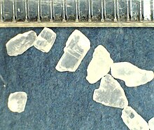
Simple ways in which the microscope can be used is a comparison of salt crystals, such as sea salt and table salt. A common millimeter scale at the tops of the micrographs show the smaller size of the cubic table salt crystals. The good depth of field available is shown by USB micrographs of a Salvia officinalis or Sage leaf.
Such devices are useful in Forensic engineering where large fracture surfaces need direct examination, an application where conventional light microscopes are restricted in use. They are normally handheld for this application, but can also be mounted in a small stand).
USB microscopes are also being used in crime scene investigation units. Many USB microscopes can view objects from a short distance, allowing sensitive crime scene evidence to be investigated without actually contaminating the evidence in any way.
They also find use in medical application such as ENT examinations.[2]
Endoscopy
Related devices include a USB endoscope, where the digital camera is fitted to a long length of cable and enables the camera to be used to inspect cavities which are otherwise difficult to examine (such as car engine interiors, pipe interiors, sewers and so on). As with the microscope, the cable is fitted with a USB plug to engage with the PC. Alternatively, a simple USB device can be fitted to a conventional endoscope. Endoscopes with a small screen are also available, allowing the user to see the hidden scene directly without the use of a laptop.
Since this area of technology is still developing very rapidly, further design and technical improvements as well as lower prices may be expected in the near future (in February 2016, there were units costing less than $10 offered on internet sites). The software used for image manipulation already offers big improvements in capability, allowing the digital images to be cropped and brightness and image contrastchanged as needed, for example. Accessories such as polarizers are expensive but allow extra control of unwanted specular reflections from the subject, for example.
Effective magnification
The precise magnification is determined by the working distance between the camera and the object, and good supports are needed to control the image, especially at higher magnifications. The magnifying abilities of these instruments are often overstated; typically offering 200× magnification, this claim is based usually on 25× to 30× actual magnification which is then further magnified by the expansion of the image by display on the screen. High magnifications are available only if the camera has a high resolution, so the image from a 5-megapixelcamera can be enlarged to a greater extent than that from a 2-megapixel camera, for example.

 ENGLISH
 中文简体
中文简体


